
PLANTS | ANIMALS | ECOLOGY | FUNGI | GEOLOGY | GARDENING | TOOLS
Excerpts from Jim Conrad's
Naturalist Newsletter
from the January 20, 2013 Newsletter issued from the valley of the Dry Frio River in northern Uvalde County, southwestern Texas, on the southern border of the Edwards Plateau; elevation ~1750m (~5750 ft); N29.62°, W99.86°; USA
EASTERN SPECKLED SHIELD LICHEN
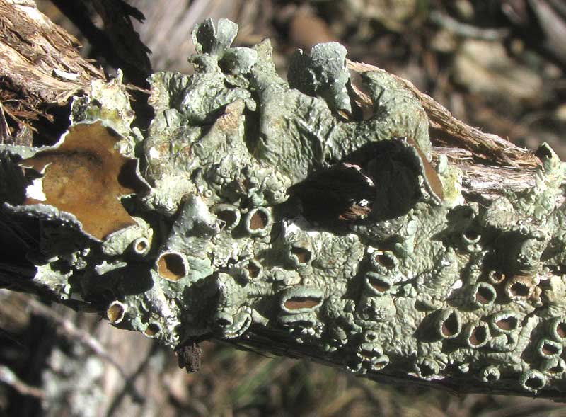
Probably the most abundant and conspicuous lichen here -- a foliose one commonly seen encrusting the bark of tree limbs, fallen twigs, and sometimes even open ground next to roads -- is the one shown above.
That's PUNCTELIA BOLLIANA, sometimes referred to as the Eastern Speckled Shield Lichen. The species is distributed in the central US from here in southwestern Texas north to North Dakota, is absent from most of the US Southeast, but present in the Appalachians and most northeastern states and adjacent Canada. The term "shield lichen" is applied to numerous foliose lichens with the general appearance of this one.
An important field mark for the Eastern Speckled Shield Lichen is the presence of many large, bowl-shaped growths scattered across the lichen's body, or thallus, with brown inner surfaces. These structures are apothecia. A side view of some apothecia displaying their bowl shape appears below:
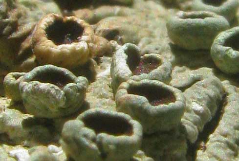
Apothecia are reproductive structures produced by the fungal component of the lichen, and of course lichens are "composite organisms" composed of a fungus and an alga and/or a cyanobacterium. The apothecium's inner surface is covered with innumerable, microscopic, tubelike affairs called asci (singular ascus) stacked next to one another and containing spores (ascospores), which escape from the tubes' tops. When the fungal spores land in a good spot they germinate, their mycelium wrap around an appropriate alga or cyanobacterium cell, and begin growing into a lichen.
Having access to a microscope, I wondered if I could see the layer of asci covering the interior surface of one of this lichen's apothecia. Maybe I could have with the proper slide-preparation equipment, but with just a knife and tweezers I couldn't because the ascus layer was so exceedingly thin. However, while fiddling with the lichen I noticed that its flat thallus surface was heavily speckled with microscopic, dark dots, as shown below:
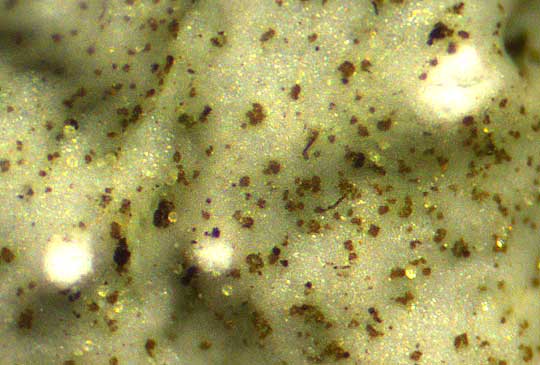
These black dots must be the speckles making this a speckled shield lichen. A little browsing on the internet revealed what the speckles were.
They're pycnidia -- singular pycnidium. Pycnidia normally are tiny, spherical or upside-down-pear-shaped (obpyriform) structures, partly or entirely sunk into the lichen's thallus, with their internal cavities lined with conidiophores, which are tips of the fungus's hyphae. The ends of these conidiophores break off forming dustlike specks, or "pycnidiospores," which then escape from the pycnidium through an opening in the pycnidium's top. A pycnidiospore can help the lichen reproduce sexually by acting as a male germ cell that unites with a female germ cell to start a new fungus, or it can accomplish asexual reproduction by landing somewhere where it can germinate a hypha that will combine with appropriate alga and/or a cyanobacterium cells and grow into a new lichen. It's as if your fingertip should break off and grow into a full-size copy of you.